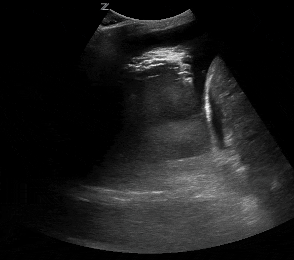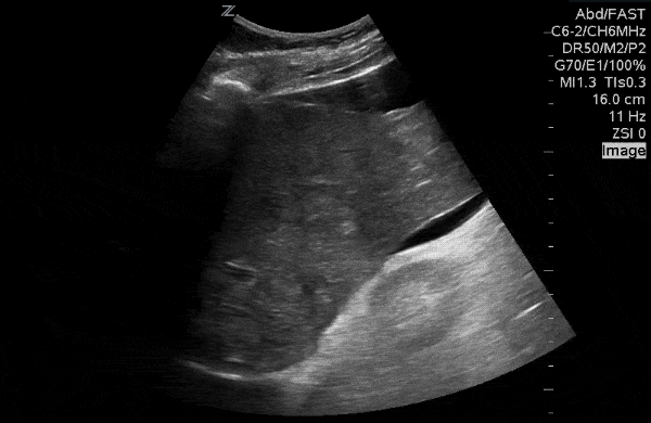
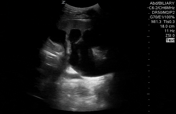
63yo M with fatigue and weight loss, generalized abdominal pain.
ANSWER
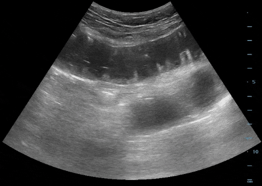
LIVER METS WITH SURROUNDING ASCITES. "Leopard-skin" appearance of the liver parenchyma. Determined to be colon cancer primary.

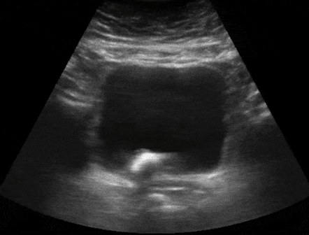
Renal ultrasound in setting of urinary retention.
ANSWER
SEVERE HYDRONEPHROSIS. Severe hydronephrosis in a patient with chronic urinary retention. Near-effacement of the cortex of the kidney.

80yo M with bladder pain.

ANSWER
BLADDER STONE. Note the echogenic stone with shadowing. The stone was freely mobile with changing patient position.

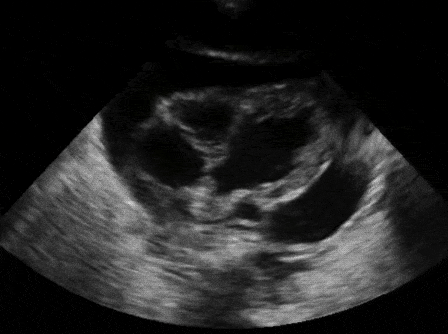
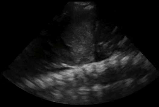
4yo with recent URI and shortness of breath.
ANSWER
CARDIAC TAMPONADE. Note the diastolic collapse of the RA and RV. RA collapse is described as "late diastolic" and RV collapse is "early diastolic".

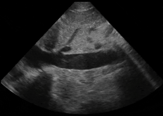
4yo s/p "accidental" stab wound to chest.
ANSWER
HEMOTHORAX. Note the "spine sign" above the diaphragm. There is a combination of clotted blood (homogeneously echogenic) and fresh blood (jet black). The lung appears to the left of the screen.

Describe the IVC.

ANSWER
LARGE, MINIMAL RESPIRATORY VARIATION. This "plethoric" IVC is suggestive of a volume overload state; at the least, you would assume this patient is not "volume tolerant" if managing hypotension/hypoperfusion.

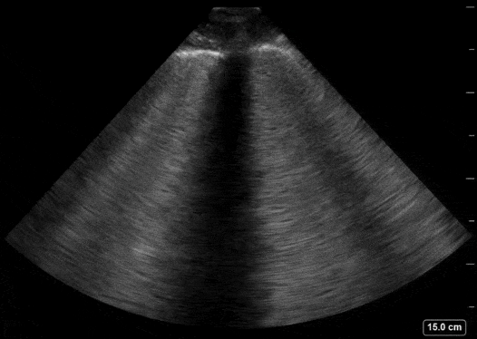
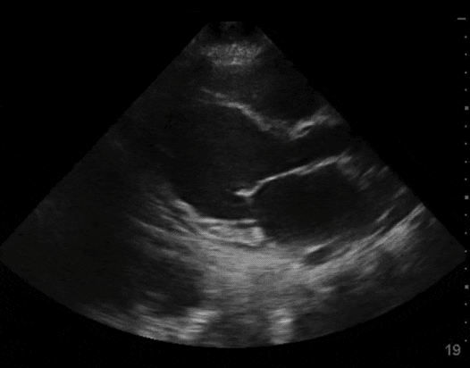
60yo M with progressive dyspnea and leg swelling.
ANSWER
PULMONARY EDEMA. Diffuse B-line pattern suggestive of pulmonary edema. Interstitial fluid produces the extensive comet tails that have a "flashlight on a foggy night" appearance.

Categorize the ejection fraction.

ANSWER
EF < 30%. Note the poor excursion of the septal leaflet of the mitral valve.

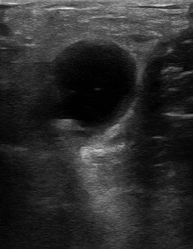
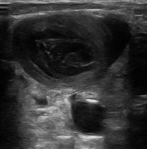
40yo F with calf pain and swelling. Imaging from the popliteal fossa.
ANSWER
BAKER'S CYST. Non-ruptured baker's cyst with the adjacent medial head of the gastrocnemius muscle (to the right of the fluid collection).

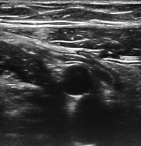
Neck swelling and minimal pain to palpation. Anterior neck evaluation.
ANSWER
IJ THROMBUS. Large internal jugular vein thrombus with carotid artery in the far field.

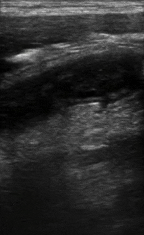
6yo M with RLQ pain. Operating room or home?
ANSWER
HOME. This is a normal appearing appendix draped over the psoas muscle (left screen) and the iliac vessels (center screen). It is tubular, blind-ended, and does not peristalse.

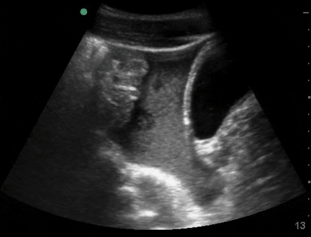
4yo with RLQ pain. Operating room or home?
ANSWER
OPERATING ROOM. This is a dilated, non-compressible appendix = appendicitis. It measured at 9mm in diameter (>6-7mm is PATHOLOGIC).

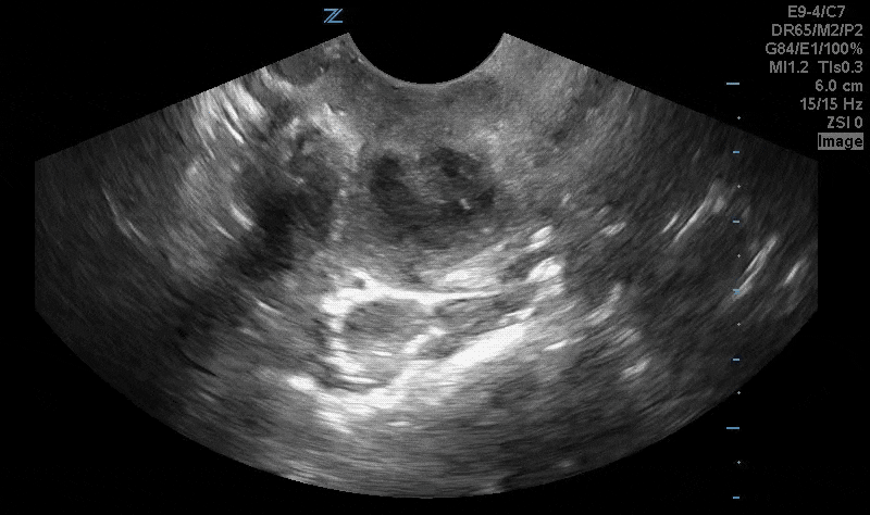
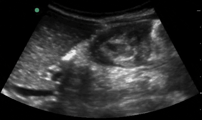
12yo with a handlebar injury. Pelvic view in sagittal. Concerned?
ANSWER
YES. The slightly echogenic material between the bladder (right) and bowel (left) is intra-peritoneal blood. While initially jet-black, free fluid from trauma can quickly coagulate and appear echogenic.

14mo F with back arching and fussiness.
ANSWER
ILEOCOLIC INTUSSUSCEPTION. Rounded structure next to the medial edge of the liver is the intussusception in short-axis. It is composed of the intussusceptum (inner bowel) which telescopes into the intussuscipiens (outer bowel).

2yo with cough and fever. What is the sonographic finding?
ANSWER
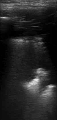
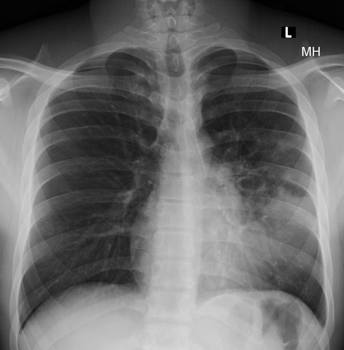
"SHRED SIGN". This refers to the irregular/uneven pleural edge due to sub-pleural consolidation. This indicates an alveolar process (pneumonia). The patient had the diagnosis confirmed on follow-up Xray.
Stacks Image 265

24yo college student with a sore throat and enlarged tonsils. Is an aspiration indicated?
ANSWER
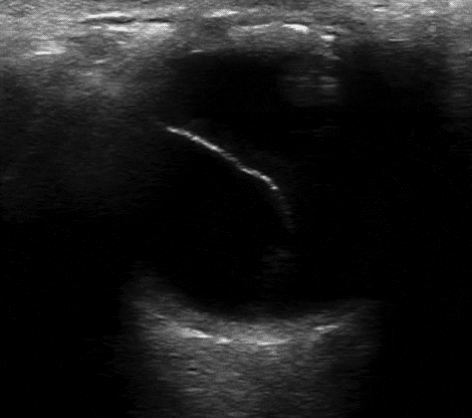
YES, THERE IS A PERI-TONSILLAR ABSCESS. Note the hypoechoic debris (center of screen) indicating an abscess.

Sudden, painless vision loss.

ANSWER
RETINAL DETACHMENT. Note the echogenic flap/line pulled away from the retina.

Decreased breath sounds at the lung base. Explanation?
ANSWER
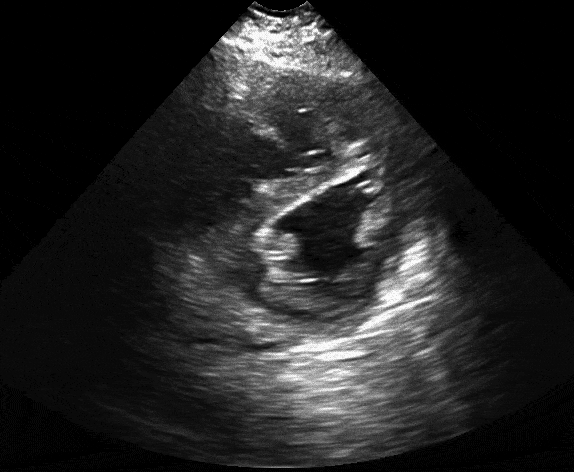
PLEURAL EFFUSION. Atelectatic lung is visualized floating in a pleural effusion, with spine visible deep to the fluid collection ("spine sign").

Parasternal short axis with a pathologic finding.
ANSWER
PARADOXICAL SEPTAL MOVEMENT. Right heart strain (RV enlargement) causing paradoxical septal movement into the LV. Appearance of a "D" shape during diastole.

Generalized abdominal pain. Frequent visits to the ED for gastroparesis.

ANSWER
SBO. Dilated loops of small bowel with "back-and-forth" or "to-and-fro" peristalsis, consistent with SBO.