Pre-Test 1
This pre-test is only to gauge your ultrasound knowledge prior to the U/S rotation.
No books or other study aids, please.
Multiple choice questions are self-explanatory. Choose the best response.
Some questions have corresponding pictures and/or U/S video clips (designated by a “*”). For the videos, click on the link that follows, which directs you to a separate web page. This page houses ALL of the videos, in order, corresponding to the questions.
Click on the “Pre-test answer sheet” link below and print out the answer sheet. You will need to use this to document your answers to the questions on the test. Enter the password below for the pre-test answer key. If you need the password, go to the Contact page and email me your request.
Good luck.
1. All of the following are true about ultrasound waves EXCEPT:
2. Attenuation is:
- Velocity of propagation is better in water than in air
- Higher frequency increases velocity
- The equation is: Velocity= Frequency X Wavelength
- Frequency is the number of cycles per second
- All of the above are correct
2. Attenuation is:
- The loss of signal energy as it passes through tissue
- The resistance to the propagation of sound
- The ability of the sound waves to discriminate between two different objects
- Anechoic signal caused by failure of the ultrasound beam to pass through an object
- None of the above
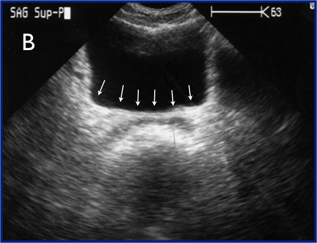
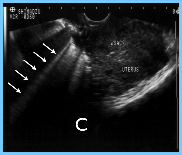
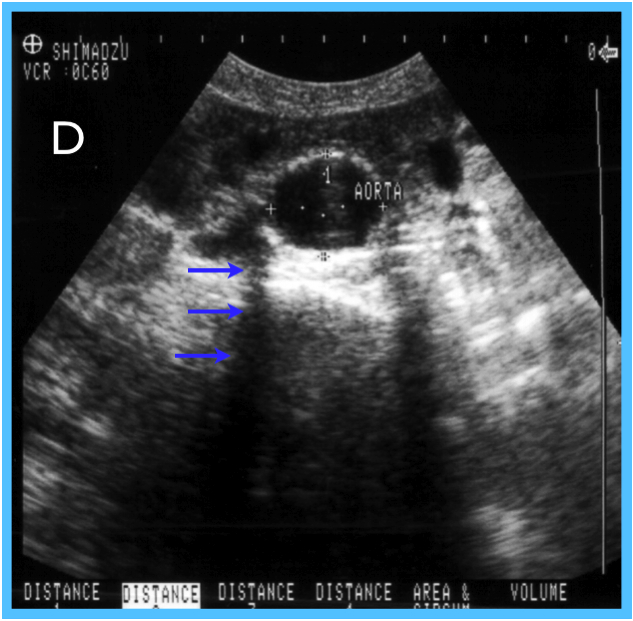
3. Identify the specific artifacts from the accompanying video and still images below:
3a
3b
3c
3d
4. True / False: The FAST exam readily identifies around 50 mL of intraperitoneal blood.
5. Name the four windows to evaluate for “free fluid”, circling the most sensitive of these locations for the identification of free fluid in a FAST exam:
- __
- __
- __
- __
6. In the Pneumothorax study with ultrasound:
- Between two rib shadows, there is a notable echogenic line composed of the visceral and parietal pleura
- The transducer is placed longitudinally (pointed cranially) in the midclavicular line
- The transducer is moved inferiorly in a systematic fashion
- A and C
- All of the above
- None of the above
7. Arrange the following sonographic findings based on the order in which they appear during pregnancy, ordering first (1) to last (5):
___Fetal pole
___Double decidual sign
___Fetal heart tones
___Yolk sac
___Thickened endometrium
8. What is the primary objective in 1st Trimester OB scans? ________________
___Fetal pole
___Double decidual sign
___Fetal heart tones
___Yolk sac
___Thickened endometrium
8. What is the primary objective in 1st Trimester OB scans? ________________
9. The aorta:
10. True / False: The aorta diameter should be measured in transverse from inside wall to inside wall.
11. In soft tissue ultrasound:
- Is approximately 2cm as it enters the abdomen and then gradually increases in size distally
- Gives off, in the following order, the celiac trunk, the superior mesenteric artery, the inferior gastric artery, the inferior mesenteric artery, and the renal arteries
- Bifurcates into the common iliac arteries at approximately the level of the umbilicus (L4 level)
- B and C
- All of the above
10. True / False: The aorta diameter should be measured in transverse from inside wall to inside wall.
11. In soft tissue ultrasound:
- Fascial planes are hyperechoic
- Muscle has a characteristic striated appearance
- Lymph nodes have echogenic centers with hypoechoic rims
- A and B
- All of the above
12. Sonographically, arteries tend to be:
- Pulsatile
- Collapsible
- Thin-walled
- All of the above
- None of the above
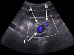
13. Identify the three structures in the Portal triad from the image to the right:
A)
B)
C)
A)
B)
C)
14. Watch the ultrasound clip of the kidney and classify the renal cyst as either simple or complex (circle your answer):
15. Name the cardiac view and the labeled structures in the video to the right:
View: ____
A)
B)
C)
D)
View: ____
A)
B)
C)
D)
16. True / False: The IVC is dilated in tamponade.
17. From the video to the right, identify the cardiac view, the diagnosis (pathology), and 2 ultrasonographic findings that support your diagnosis:
View: __________________
Diagnosis: _______________
1)
2)
View: __________________
Diagnosis: _______________
1)
2)
Congratulations!! You're done!!


